CPB Mechanics of Epithelia Symposium
March 23, 1-6 pm
A half-day symposium that will bring together scientists using biophysical, molecular and theoretical approaches to study the dynamics and mechanics of epithelial morphogenesis.
Scientific organizer - Stephanie Höhn
Programme (GMT time)
Abstracts: scroll down or click the speakers names
Poster competition information: here
Join our Slack workspace: here
13:00 – 13:05 – Welcome
Part I
13:05 – 13:30 – Katja Röper Mechanisms and mechanics of tube morphogenesis
MRC LMB Cambridge, UK
13:30 – 13:45 – Natalie Dye Self-organized patterning of cell morphology via mechanosensitive feedback
MPI-CBG Dresden, Germany
13:45 – 14:10 – Stephan Grill Tissue mechanics and the cell death decision in the germline
MPI-CBG Dresden, Germany
14:10 – 14:35 – Alexandre Kabla Rheology and curvature of epithelial monolayers
Dept. Engineering University of Cambridge, UK
14:35 – 14:50 – Break 1
Part II
14:50 – 15:05 – Akanksha Jain Regionalized tissue morphogenesis in insect embryos and human cerebral organoids
ETH Zürich D-BSSE, Switzerland
15:05 – 15:30 – Stephanie Höhn
DAMTP - University of Cambridge, UK
15:30 – 15:55 – Guillaume Salbreux Cellular rules behind tissue growth
Francis Crick Institute, London, UK
15:55 – 16:20 – Caren Norden Matrix topology guides collective cell migration in optic cup formation
Instituto Gulbenkian Ciencia, Portugal
16:20 – 16:35 – Break 2
Part III
16:35 – 16:50 – Nicholas Noll Variational stress inference: a robust method to measure tissue mechanics
Kavli Institute for Theoretical Physics, Santa Barbara, USA
16:55 – 17:15 – Lance Davidson Mechanical discordance and jamming during neurulation
University of Pittsburgh, USA
17:15 – 17:40 – Kinjal Dasbiswas A mechanochemical wrinkle on tissue shape
University California - Merced, USA
17:40 – 18:00 – Future directions and closing
Abstracts
Katja Röper - Mechanisms and mechanics of tube morphogenesis
We study the dynamic behaviour of epithelial sheets of cells during organ formation, in particular during the formation of tubular organs, using the formation of the tubes of the salivary glands in the Drosophila embryo as a main model system. These tubes form through a process of budding, and we have uncovered that cell behaviours across the tissue primordium, the placode, are highly patterned during the initial formation of the tube from a flat epithelial sheet. Within the apical domain, isotropic constriction near the invagination point combines with polarised cell intercalation away from the invagination point. In 3D, this is due to strong wedging of cells near the pit, as well as tilting towards it, and interleaving of cells across the tissue. I will discuss these findings in the light of the analyses of mutants that fail proper tube formation and that allow us to dissect the contribution of different behaviours, switching of cells between behaviours, as well as their potential mechanical interplay. Group website
Natalie Dye - Self-organized patterning of cell morphology via mechanosensitive feedback
Tissue organization is often characterized by specific patterns of cell morphology. How such patterns emerge in developing tissues is a fundamental open question. In this talk, I will present our group's recent investigation into the emergence of tissue-scale patterns of cell shape and mechanical tissue stress in the Drosophila wing imaginal disc during larval development. Using quantitative analysis of the cellular dynamics, we revealed a pattern of radially oriented cell rearrangements that is coupled to the buildup of tangential cell elongation. I will present our continuum theory showing that this pattern of cell morphology and tissue stress can arise via self-organization of a mechanical feedback that couples cell polarity to active cell rearrangements. The predictions of this model are supported by knockdown of MyoVI, a component of mechanosensitive feedback. Our work reveals a novel mechanism for the emergence of cellular patterns in morphogenesis. Group website
Stephan Grill - Tissue mechanics and the cell death decision in the germline
Oocytes transmit genetic information to the next generation. They are large cells, providing sufficient cellular material to develop into an embryo upon fertilization. As germ cells mature into oocytes some germ cells grow, typically at the expense of others that undergo cell death. The associated exchange of material is facilitated by a syncytial structure with a shared cytoplasm. How germ cells are selected to live or die out of a seemingly homogeneous population of healthy cells remains unclear. I will present evidence that in the gonad of the nematode Caenorhabditis elegans, this cell fate decision is mechanical and related to tissue hydraulics. An analysis of germ cell volumes and cytoplasmic fluxes reveals a transition in germline hydraulics at approximately 60% gonad length, involving an inversion of the pressure difference between germ cells and the shared cytoplasmic pool. A theoretical description of gonad mechanics identifies a hydraulic instability that amplifies volume differences and causes some germ cells to grow and others to shrink, a phenomenon that is related to the well known two-balloon instability. Shrinking germ cells are then extruded and die, as we demonstrate by artificially reducing germ cell volumes via thermoviscous pumping. Our work reveals a hydraulic symmetry breaking transition that is central to the life and death cell fate decision in the nematode germline. Group website
Alexandre Kabla - Rheology and curvature of epithelial monolayers
Cell migration and cell mechanics play a crucial role in a number of key biological processes, such as embryo development or cancer metastasis. It is therefore important to characterise the material properties of cells and tissues and the way they mechanically interact with their environment. In this talk, I will present recent work to address these questions at the tissue level. Experimental studies on the mechanical response of in-vitro epithelial monolayers show that the material exhibits a strong time-dependant response over a broad range of time-scales. In this situation, it is challenging to capture the response of the system with a few parameters without losing some of the material’s characteristic features. Rheological models based on fractional calculus are effective empirical tools to summarize such complex data and highlight similarities across a broad range of systems. The polarity of the tissue also induces differential contractility between the apical and basal side of the monolayer leading to spontaneous curvature and the curling of the monolayer along free edges. Group website
Akanksha Jain - Regionalized tissue morphogenesis in insect embryos and human cerebral organoids
Organisms adopt diverse developmental strategies to sculpt tissues into characteristic 3D shapes with molecularly and mechanically distinct regions. We are using longterm lightsheet imaging, quantitative analysis and spatial transcriptomics to study different aspects of regionalised tissue behaviours exhibited during embryogenesis in insects and self-patterning of cerebral organoids. Extra embryonic tissue (serosa) in the short germ insect Tribolium castaneum undergoes dramatic epiboly like expansion and ventral window closure to form a protective shell around the growing embryo. This creates mechanical stress on the dorsally expanding tissue which exhibits contractility and increasingly resists expansion during serosa window closure. To accomplish a seamless serosa window closure, the beetle serosa undergoes a regional tissue fluidisation and structural transition as the epithelium expands. This behaviour requires the contractile activity of a supracellular actomyosin cable that forms at the serosa-embryo tissue boundary and promotes local cell rearrangements. Next, we are using longterm live imaging coupled with lineage tracking and barcoding to study how lineages are established during cerebral organoid regionalization. Cerebral organoids self-pattern and generate diverse luminal regions closely resembling the foetal neocortex, in cell type composition as well as structural organisation. Our results show that cerebral organoids exhibit early commitment to specific brain regions followed by local proliferation and clonal expansion in a single lumen. Personal page
Stephanie Höhn - Mechanics and elasticity of dynamic cellular monolayers
Stephanie S.M.H. Höhn, Pierre A. Haas, and Raymond E. Goldstein. Living tissues are intelligent materials that can change their mechanical properties while they develop. In spite of extensive studies, we are only just beginning to understand these dynamic properties and their role in tissue development. Although many tissues are known to exhibit visco-elastic behaviour, it is unclear which properties dominate three-dimensional shape changes of cellular monolayers, such as epithelia. Here we explore the mechanics of an initially spherical cellular monolayer undergoing dramatic topological changes including invagination and complete inversion [1]. These global deformations are driven by several waves of cell shape changes causing local bending, contraction and expansion [2, 3]. A combination of advanced imaging, laser ablation experiments and computational analyses is used to determine internal stresses and elastic behaviour of the dynamic cell sheet [4]. [1] Höhn S and Hallmann A. BMC Biology 9, 89 (2011). [2] Höhn S, Honerkamp-Smith AR, Haas PA, Khuc Trong P, and Goldstein RE. Physical Review Letters 114, 178101 (2015). [3] Haas PA, Höhn S, Honerkamp-Smith AR, Kirkegaard JB, and Goldstein RE. PLOS Biology 16, e2005536 (2018). [4] Höhn S, Haas PA, and Goldstein [in preparation]. Personal page
Guillaume Salbreux - Cellular rules behind tissue growth
Shape changes of a biological tissue are determined by mechanical stresses acting within the tissue cells. To understand tissue morphogenesis, cellular scale processes must be related to flows and deformations occurring at the tissue scale. Here I will discuss how large-scale tissue expansion is related to cellular events during the growth of histoblast nests in the Drosophila abdomen. We find that tissue expansion is primarily driven by cell proliferation rather than area growth and that cell proliferation appears largely insensitive to geometric cues. By examining tissue mechanics throughout histoblast expansion, we uncover a decrease in extracellular matrix resistance as the histoblasts expand, which is necessary to trigger tissue expansion. Later, growth arrest occurs through stochastic transitions of dividing cells to quiescence. I will describe how oscillations in cell division rate during growth are related to neighbour couplings between cell cycle times, and how an apparent adder behaviour during the cell cycle ensures a tight control of cell area in the tissue. Group website
Caren Norden - Matrix topology guides collective cell migration in optic cup formation
Karen Soans, Carl Modes, Caren Norden. Collective migration of epithelial cells often plays crucial roles in shaping tissues during development. One prominent example of collective cell migration in morphogenesis is rim cell migration during optic cup formation. Here, cells collectively migrate from one side of the optic cup that will become retinal pigment epithelium into the neuroepithelium where these cells will take on neuro-progenitor fate. We previously showed that in zebrafish, rim migration is an active form of collective cell migration that depends on an intact underlying extracellular matrix. However, we did not yet understand how exactly cell-matrix interactions influence this process and whether the matrix plays a direct or indirect role. Investigating this question, we find that laminin forms a stable structure with different topologies that rim cells actively migrate over. Migrating rim cells display distinct focal adhesion dynamics on these different topologies. Knocking down laminin results in aberrant topologies and is leads to less directed rim cell migration suggesting their directed movement could be a form of ‘topotaxis’. We now set out to also dissect what triggers directed migration in the first place and how directionality is preserved. Group website
Nicholas Noll - Variational stress inference: a robust method to measure tissue mechanics
While cellular mechanics drives epithelial morphogenesis, quantitative understanding is impaired by the difficulty of the direct measurement of stress in-vivo. In this talk, I will introduce an approach based on a novel parametrization of epithelial tissues in terms of polygonal tilings with circular arc edges. These polygons represent the equilibrium geometry of an array of cells, allowing us to identify observable constraints on the cellular geometry that are imposed by mechanical equilibrium.
By imposing such constraints on the image analysis of epithelia, we formulate a robust algorithm that simultaneously segments cells and infers cell-scale stress over the entire tissue. Lastly, I will show the utility of our approach by comparing our predictions against the measured global pattern of myosin II during early Drosophila embryogenesis. Personal webpage
Lance Davidson - Mechanical discordance and jamming during neurulation
Early neurulation movements in vertebrates coordinately position multiple cell layers in anticipation of the folding and fusion events that form the neural tube and the central nervous system. In this presentation I discuss findings from recent live imaging, computational simulation, and embryological manipulations of the developing Xenopus frog. Using both off-the-self and custom image analysis tools, we map T1 transitions and compare cell- and tissue-strain rates during a rapid phase of convergent extension that shapes the neural plate. We compare these morphological changes in the neural epithelium with a fully passive computational model of convergent extension. Live cell data from free and physically-constrained neural plate explants suggest regionalization of jammed and unjammed cells. We propose the spatial distribution of jammed tissues reflect patterns of passive and active cell rearrangement behaviors across the neural plate. The goal of these studies is to identify signatures of active cell rearrangement and determine the role of passive cell responses in epithelial morphogenesis. Group website
Kinjal Dasbiswas - A mechano-chemical wrinkle on tissue shape
Biological processes including tissue morphogenesis are inherently mechano-chemical. Epithelial tissue sheets require coordinated cell shape changes regulated by global patterning of mechanical forces by biochemical gradients. Inspired by such pattern formation, we propose a minimal mechanochemical model based on the notion that cell shape changes are induced by diffusible biomolecules that influence tissue contractility in a concentration-dependent manner - and whose concentration is in turn affected by the macroscopic tissue shape. After theoretically exploring model cell shape profiles induced by concentration gradients, we perform computational simulations of thin shell elastic dynamics to reveal the propagating chemical and three-dimensional deformation patterns. The latter exhibit a wrinkling pattern arising from a sequence of buckling instabilities that can be understood from the theory of elastic shells. Depending on the concentration threshold that actuates cell shape change, we find qualitatively different patterns. These mechanochemically coupled patterning dynamics are distinct from those driven by purely mechanical or purely chemical factors and show long-range propagation even without chemical diffusion. Group website
Poster Session
via Slack
To participate in the poster session please submit your poster (ppt or pdf) and abstract (max 250 words) by email to admin@physbiol.cam.ac.uk until the 19th March 5 pm.
22th - 24th March
Slack channels will be open for discussion from 9 am on the 22th March until 5 pm on the 24th March for both poster session presenters and invited speakers. Prior to the event we will create a slack #channel for your poster but you are responsible to upload the poster file yourself.
Recommendation for posters:
Although any dimensions can be used, remember that attendees will be viewing posters on their computer screens.
We recommend:
1) Posters to be readable without zoom when displayed at full-screen width. (try arranging information blocks that can read horizontal instead of the traditional vertical arrangement)
2) You upload an optional audio/video walkthrough of your poster presentation (no longer than 10 minutes), which will act as a guided presentation for attendees to view or listen to while viewing your poster.
Prior to the event we will create a slack #channel for your poster but you are responsible to upload the poster file yourself.
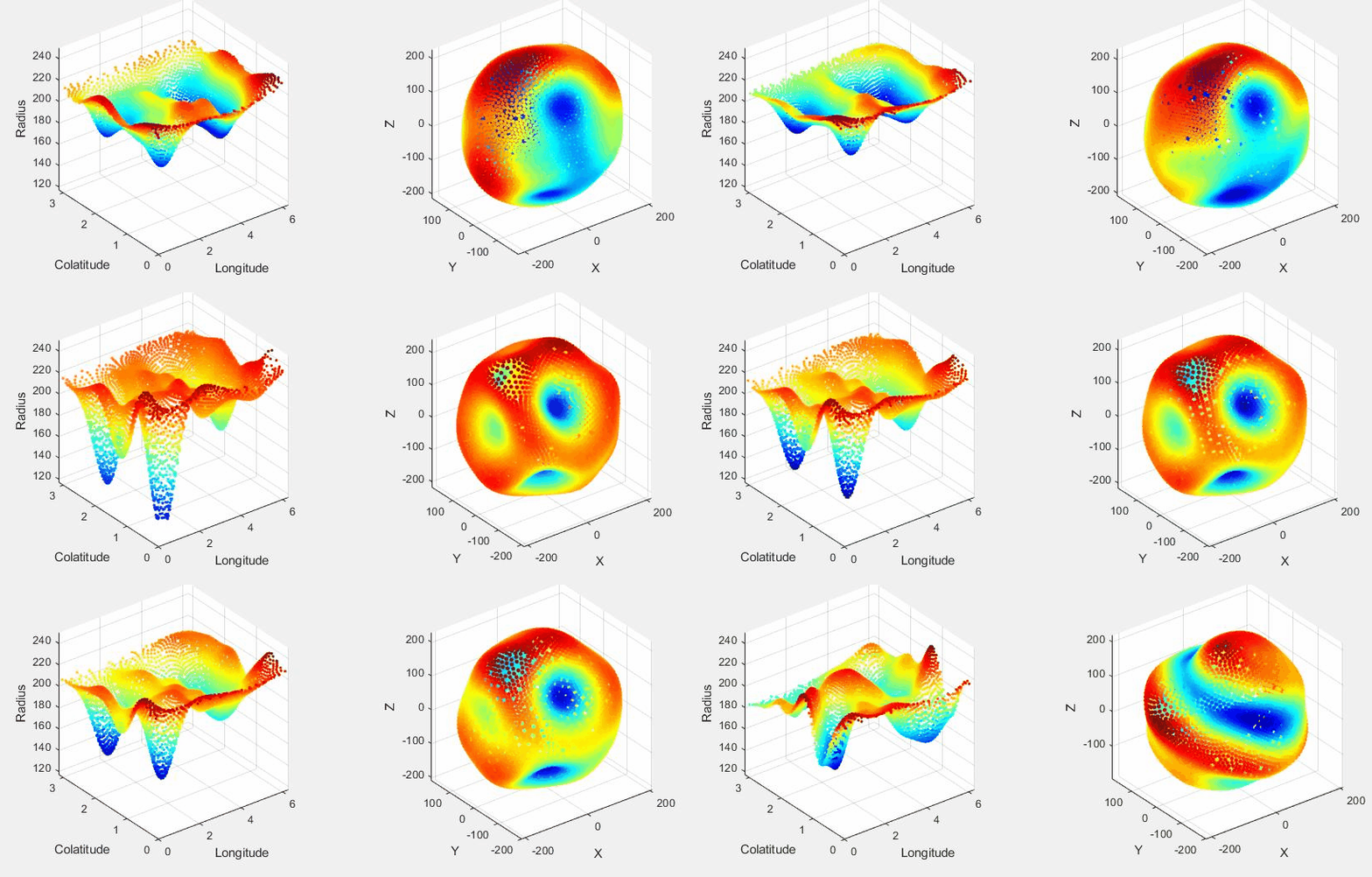
Figure legend: Shape fluctuations preceding embryonic inversion in Volvox globator by Stephanie Höhn


